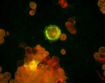
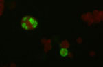
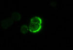
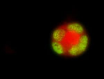
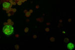
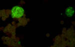
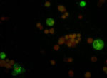
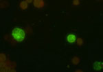
Antibodies to Epstein-Barr Virus Proteins
- anti-EBV Nuclear Antigen-1 (EBNA-1)
- Mouse monoclonal IgG2
- Recommended for Western blotting, immunoprecipitation, immunohistochemistry
- Stains formalin fixed paraffin-embedded tissue sections after antigen retrieval
(see recommended protocols)
- Epitope corresponding to aminoacids 610-640 mapping at the carboxyl terminus
of EBNA-1 protein
- Using immunostaining the antigen is detected in the nucleus by this antibody
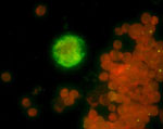
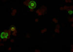
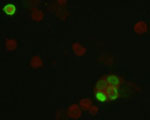
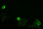
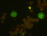
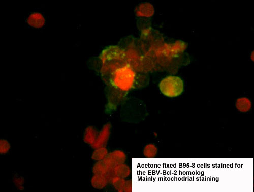
- anti-EBV Early Antigen-R (EA-R) Bcl-2 homologue
- Mouse monoclonal IgG1
- Recommended for Western blotting, immunoprecipitation, immunohistochemistry
- Stains formalin fixed paraffin-embedded tissue sections after antigen retrieval
(see recommended protocols)
- Using immunostaining the antigen is detected in the cytoplasm by this antibody
(staining pattern changes with time in the life cycle of the virus)
 Click on the pictures
- anti-EBV interleukin-10 (v-IL-10) IL-10 homologue
- Mouse monoclonal IgG1
- Recommended for Western blotting, immunoprecipitation, immunohistochemistry
- Stains formalin fixed paraffin-embedded tissue sections after antigen retrieval
(see recommended protocols)
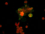
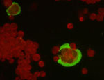
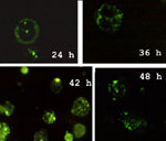
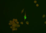
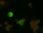
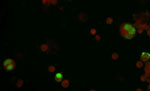
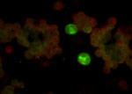
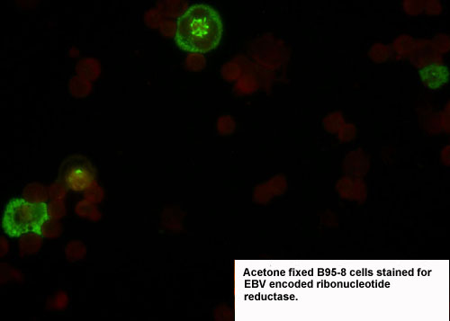
- anti-EBV Early Antigen-R (EA-R) ribonucleotide reductase large subunit
- Mouse monoclonal IgG2
- Recommended for Western blotting, immunoprecipitation, immunohistochemistry
- Ribonuclease activity is not inhibited by this antibody in in vitro assay
- Stains formalin fixed paraffin-embedded tissue sections after antigen retrieval
(see recommended protocols)
- Using immunostaining the antigen is detected in the cytoplasm by this antibody
(staining pattern changes with time in the life cycle of the virus see also
gallery)
 Click
on the pictures
Click
on the pictures
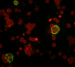
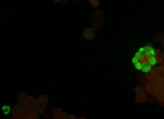
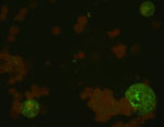
- anti-EBV Early Antigen-R (EA-R) ribonucleotide reductase large subunit
- Mouse monoclonal IgG1
- Recommended for immunoprecipitation, immunohistochemistry
- Stains formalin fixed paraffin-embedded tissue sections after antigen retrieval
(see recommended protocols)
- This antibody does not recognize the antigen on Westernblotts
- Ribonuclease activity is inhibited by this antibody in in vitro assay
- Recognized epitope is different from the previous antibody
Using immunostaining the antigen is detected in the cytoplasm by this antibody
- anti-EBV Early Antigen-R (EA-R) ribonucleotide reductase large subunit
- Mouse monoclonal IgG1
- Recommended for immunoprecipitation, immunohistochemistry
- Stains formalin fixed paraffin-embedded tissue sections after antigen retrieval
(see recommended protocols)
- This antibody does not recognize the antigen on Westernblotts
- Recognized epitope is different from the previous two antibodies
Using immunostaining the antigen is detected in the cytoplasm by this antibody
 Click on the picture
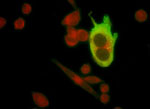
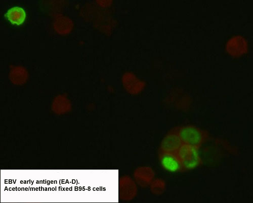
- anti-EBV Early Antigen-D (EA-D) p47-53
- Mouse monoclonal IgG2
- Recommended for Western blotting, immunoprecipitation, immunohistochemistry
- Stains formalin fixed paraffin-embedded tissue sections after antigen retrieval
(see recommended protocols)
- Using immunostaining the antigen is detected in the nucleus and cytoplasm
by this antibody
- Epitope is at the carboxyl terminus of the protein
 Click on the pictures
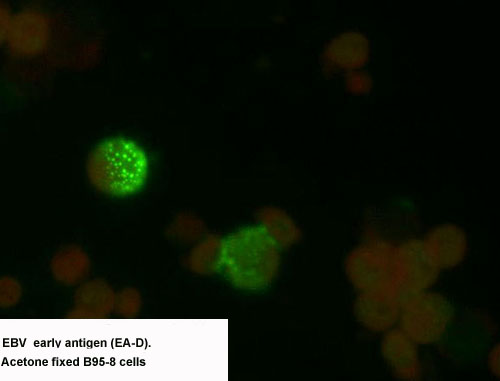
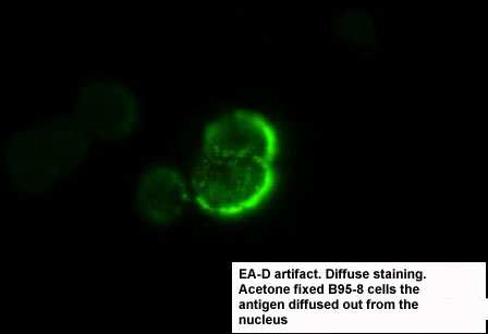
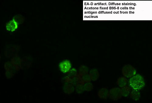
- anti-EBV Early Antigen-D (EA-D) p47-53
- Mouse monoclonal IgG2
- Recommended for Western blotting, immunoprecipitation, immunohistochemistry
- Stains formalin fixed paraffin-embedded tissue sections after antigen retrieval
(see recommended protocols)
- Using immunostaining the antigen is detected in the nucleus and cytoplasm
by this antibody (occasionally the antigen can diffuse out from the nucleus
and the staining is mainly cytoplasmic {last two pictures. This happens if
the drying of the cells on the slides was to slow)
- Epitope is at the middle part of the protein
-
 Click on the pictures
- anti-EBV Viral Capsid Antigen (VCA) p 140
- Mouse monoclonal IgG2
- Recommended for immunoprecipitation, immunohistochemistry
- Stains formalin fixed paraffin-embedded tissue sections after antigen retrieval
(see recommended protocols)
- This antibody does not recognize the antigen on Westernblotts
- Using immunostaining the antigen is detected in the nucleus by this antibody
-

 Click on the pictures
- anti-EBV Viral Capsid Antigen (VCA) p 140
- Mouse monoclonal IgG2
- Recommended for immunoprecipitation, immunohistochemistry
- Stains formalin fixed paraffin-embedded tissue sections after
- antigen retrieval (see recommended protocols)
- This antibody recognizes the antigen on Westernblotts but the signal is
weak
- Recognized epitope is different from the previous antibody
Using immunostaining the antigen is detected in the nucleus by this antibody
-
 Click on the pictures
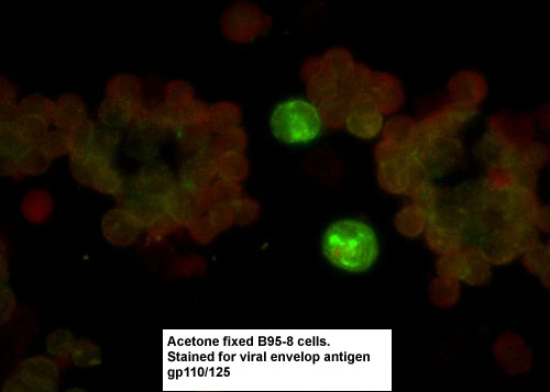
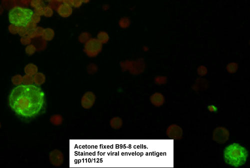
- anti-EBV Envelope Protein (VCA) gp 110-120
- Mouse monoclonal IgG2
- Recommended for Western blotting, immunoprecipitation, immunohistochemistry
- Stains formalin fixed paraffin-embedded tissue sections after antigen retrieval
(see recommended protocols)
- Using immunostaining the antigen is detected in the cytoplasm by this antibody
-
 Click on the pictures
- anti-EBV Envelope Protein (VCA) GP 110-120
- Mouse monoclonal IgG2
- Recommended for Western blotting, immunoprecipitation, immunohistochemistry
- Stains formalin fixed paraffin-embedded tissue sections after antigen retrieval
(see recommended protocols)
- Recognized epitope is different from the previous antibody
Using immunostaining the antigen is detected in the cytoplasm by this antibody
-
 Click
on the pictures
Click
on the pictures
- anti-EBV Envelope Protein (VCA) GP 110-120
- Mouse monoclonal IgG2
- Recommended for Western blotting, immunoprecipitation, immunohistochemistry
- Stains formalin fixed paraffin-embedded tissue sections after antigen retrieval
(see recommended protocols)
- Recognized epitope is different from the previous two antibodies
Using immunostaining the antigen is detected in the cytoplasm by this antibody
- anti-EBV Membrane Antigen (MA) gp220-350
- Mouse monoclonal IgG1
- Recommended for immunoprecipitation, immunohistochemistry
- Stains formalin fixed paraffin-embedded tissue sections after antigen retrieval
(see recommended protocols)
- This antibody does not recognize the antigen on Westernblotts
- Using immunostaining the antigen is detected in the cytoplasm and on the
membrane by this antibody
-
 Click on the pictures
- anti-EBV Membrane Antigen (MA) gp350
- Mouse monoclonal IgG1
- Recommended for immunoprecipitation, immunohistochemistry
- Stains formalin fixed paraffin-embedded tissue sections after antigen retrieval
(see recommended protocols)
- This antibody does not recognize the antigen on Westernblotts
- Recognized epitope is different from the previous antibody
Recognizes only A-type EBV antigen.
- Using immunostaining the antigen is detected in the cytoplasm and on the
membrane by this antibody
-
 Click on the picture
- anti-EBV Membrane Antigen (MA) gp350
- Mouse monoclonal IgG1
- Recommended for immunoprecipitation, immunohistochemistry
- Stains formalin fixed paraffin-embedded tissue sections after antigen retrieval
(see recommended protocols)
- This antibody recognizes the antigen on Westernblotts
- Recognized epitope is different from the previous two antibodies
Recognizes unglycosylated and glycosylated forms of the antigen
- Using immunostaining the antigen is detected in the cytoplasm by this antibody
Antibodies to Human Herpesvirus 6
- anti-HHV-6 Viral Capsid Antigen (VCA) p 140 (H-JG-16)
- Mouse monoclonal IgG1
- Recommended for immunoprecipitation, immunohistochemistry
- Stains formalin fixed paraffin-embedded tissue sections after antigen
retrieval (see recommended protocols)
- This antibody does not recognize the antigen on Westernblotts
- Using immunostaining the antigen is detected in the nucleus by this
antibody
- anti-HHV-6 Viral Capsid Antigen (VCA) p 140 (H-JG-20)
- Mouse monoclonal IgG1
- Recommended for immunoprecipitation, immunohistochemistry
- Stains formalin fixed paraffin-embedded tissue sections after antigen
retrieval (see recommended protocols)
- This antibody does not recognize the antigen on Westernblotts
- Using immunostaining the antigen is detected in the nucleus by this
antibody
Recognized epitope is different from the previous antibody
- anti-HHV-6 Envelope Antigen (VCA) GP 85-110 (H-AR-1)
- Mouse monoclonal IgG2
- Recommended for immunoprecipitation, immunohistochemistry and westernblotting
- Stains formalin fixed paraffin-embedded tissue sections after antigen
retrieval (see recommended protocols)
- Using immunostaining the antigen is detected in the cytoplasm and on
the membrane by this antibody
- Neutralizes virus infection in vitro
- anti-HHV-6 Envelope Antigen (VCA) GP 85-110 (H-AR-2)
- Mouse monoclonal IgG2
- Recommended for immunoprecipitation, immunohistochemistry and westernblotting
- Stains formalin fixed paraffin-embedded tissue sections after antigen
retrieval (see recommended protocols)
- Using immunostaining the antigen is detected in the cytoplasm and on
the membrane by this antibody
- Neutralizes virus infection in vitro
- Recognized epitope is different from the previous antibody
anti-HHV-6 Envelope Antigen (VCA) GP 85-110 (H-AR-4)
- Mouse monoclonal IgG2
- Recommended for immunoprecipitation, immunohistochemistry and westernblotting
- Stains formalin fixed paraffin-embedded tissue sections after antigen
retrieval (see recommended protocols)
- Using immunostaining the antigen is detected in the cytoplasm and on
the membrane by this antibody
- Neutralizes virus infection in vitro
- Recognized epitope is different from the previous two antibodies
anti-HHV-6 Envelope Antigen (VCA) GP 85-110 (H-AR-6)
- Mouse monoclonal IgG1
- Recommended for immunoprecipitation, immunohistochemistry and westernblotting
- Stains formalin fixed paraffin-embedded tissue sections after antigen retrieval
(see recommended protocols)
- Using immunostaining the antigen is detected in the cytoplasm and on the
membrane by this antibody
- Neutralizes virus infection in vitro
- Recognized epitope is different from the previous three antibodies
- anti-HHV-6 Membrane Antigen (MA) GP 90 (H-AR-8)
- Mouse monoclonal IgG2
- Recommended for immunoprecipitation, immunohistochemistry and westernblotting
- Stains formalin fixed paraffin-embedded tissue sections after antigen retrieval
(see recommended protocols)
- Using immunostaining the antigen is detected in the cytoplasm and on the
membrane by this antibody
- Partially neutralizes virus infection in vitro
Antibodies to Human Herpesvirus 7
- anti-HHV-7 Envelope Antigen (VCA) GP 90-110
- Mouse monoclonal IgG2
- Recommended for immunoprecipitation, immunohistochemistry and westernblotting
- Stains formalin fixed paraffin-embedded tissue sections after antigen
retrieval (see recommended protocols)
- Using immunostaining the antigen is detected in the cytoplasm and on
the membrane by this antibody
- Neutralizes virus infection in vitro
- Weak cross-reactivity with HHV-6 envelop protein
- Recognized epitope is different from the previous antibody
Antibodies to Cytomegalovirus
- anti-CMV tegument protein (VCA) p68
- Mouse monoclonal IgG2
- Recommended for immunoprecipitation, immunohistochemistry and westernblotting
- Stains formalin fixed paraffin-embedded tissue sections after antigen
retrieval (see recommended protocols)
- Using immunostaining the antigen is detected in the nucleus and cytoplasm
by this antibody
- anti-CMV Immediately early antigen (IEA) p72
- Mouse monoclonal IgG2
- Recommended for immunoprecipitation, immunohistochemistry and westernblotting
- Stains formalin fixed paraffin-embedded tissue sections after antigen
retrieval (see recommended protocols)
- Using immunostaining the antigen is detected in the nucleus by this
antibody
Back to Janos Luka's Home Page
Revised: June 5, 2000


 Click on the pictures
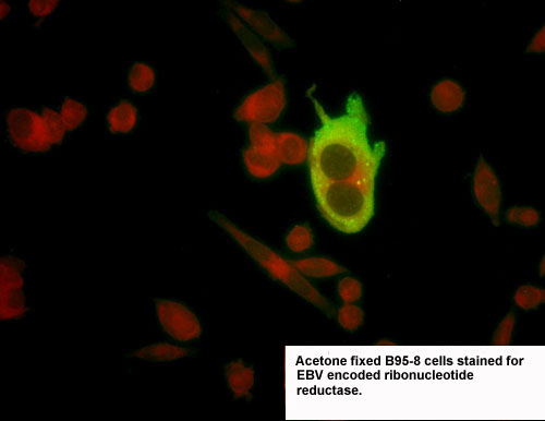

 Click on the picture
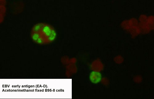
 Click on the pictures
 Click on the pictures
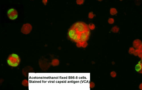
 Click on the pictures
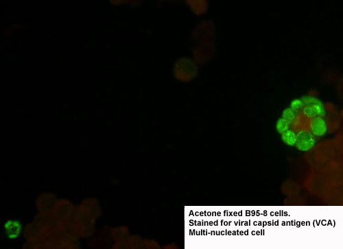
 Click on the pictures
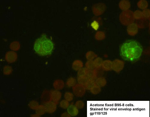
 Click on the pictures
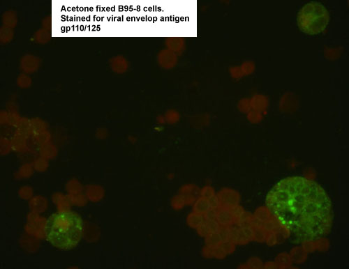
 Click
on the pictures
Click
on the pictures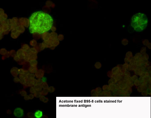
 Click on the pictures
 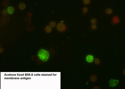 Click on the picture