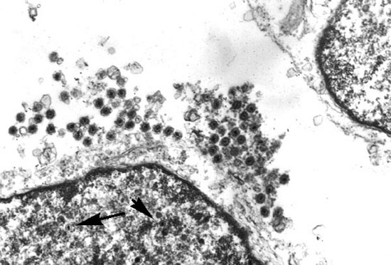
HSB-2 cell line was infected with HHV-6 (GS strain) and the infected cells were processed for electron microscopy at day 6. Arrows show HHV-6 capsids in the nucleus. Partially enveloped virus can be seen in the remains of the cytoplasm.
Copyright © 2000 Janos Luka. All rights reserved. See Term for Image use for information regarding the use of these images for education or Web Sites.